41 microscope diagram without labels
› doc › 232757022K To 12 Science Grade 7 Learners Material - Module Read and do the activities in the section on How to Use The Light Microscope before performing Activity 2. Activity 2 Investigating plant cells Objectives In this activity, you should be able to: 1. prepare a wet mount; 2. describe a plant cell observed under the light microscope; 3. stain plant cells; 4. Labeling the Parts of the Microscope | Microscope World Resources Labeling the Parts of the Microscope, This activity has been designed for use in homes and schools. Each microscope layout (both blank and the version with answers) are available as PDF downloads. You can view a more in-depth review of each part of the microscope here. Download the Label the Parts of the Microscope PDF printable version here.
alex.state.al.us › plansALEX | Alabama Learning Exchange Students will use a Venn diagram to compare lightning and static electricity. Then, students will experiment with static electricity and read nonfiction passages about lightning and lightning rods. Finally, they will apply their learning to construct a model of a lightning rod system that protects a house from a lightning-induced fire.

Microscope diagram without labels
Microscope Objective Lens | Products | Leica Microsystems Microscope Objectives. Leica Microsystems – The Ultimate in Optical Competence. For more than 170 years Leica Microsystems has designed and produced top-class objectives for a wide variety of applications in research, industry and medicine. The optics specialists at Leica Microsystems bring the highest level of experience and expertise to bear in reducing … Label the Microscope Diagram | Download Scientific Diagram - ResearchGate Download scientific diagram | Label the Microscope Diagram from publication: Laboratory Exercises in Microbiology: Discovering the Unseen World through Hands-on Investigation | Microbiology ... 7th grade Science - Microscope Diagram | Quizlet The Parts of a Microscope. 12 terms. totobear PLUS. Sets found in the same folder. Science Key terms 7th grade. 13 terms. palocastillo. 7th Grade Earth Science. 9 terms. EliseC17. 7thGrade Review - Cells/Biology. 26 terms. SolizScience TEACHER. 7th grade Science, Cell theory. 8 terms. Super1412. Other sets by this creator.
Microscope diagram without labels. en.wikipedia.org › wiki › Nuclear_envelopeNuclear envelope - Wikipedia The spindle fibers either form within the membrane, or penetrate it without tearing it apart. In other eukaryotes (animals as well as plants), the nuclear membrane must break down during the prometaphase stage of mitosis to allow the mitotic spindle fibers to access the chromosomes inside. The breakdown and reformation processes are not well ... Parts of Stereo Microscope (Dissecting microscope) – labeled diagram ... Compared to a compound microscope where the objectives attached to the nosepiece can be seen and identified individually (based on color bands and their respective labels), the objectives of a dissecting microscope are located in a cylindrical cone and, therefore, are not directly seen. For the stereo microscope that comes with multiple objective lens sets (fixed power style), the … A Study of the Microscope and its Functions With a Labeled Diagram ... To better understand the structure and function of a microscope, we need to take a look at the labeled microscope diagrams of the compound and electron microscope. These diagrams clearly explain the functioning of the microscopes along with their respective parts. Man's curiosity has led to great inventions. The microscope is one of them. Amazing 27 Things Under The Microscope With Diagrams - Microbe Notes Under a high-power microscope, the cell organelles are more differentiated and allow the observation of individual structures. Because of the affinity of the stain with the DNA and RNA of the cell, the components inside the nucleus might also be visible. 10. DNA under the microscope.
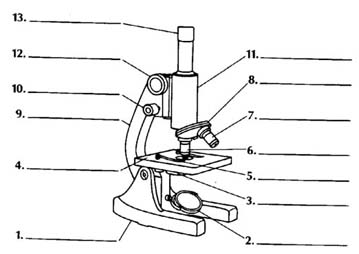
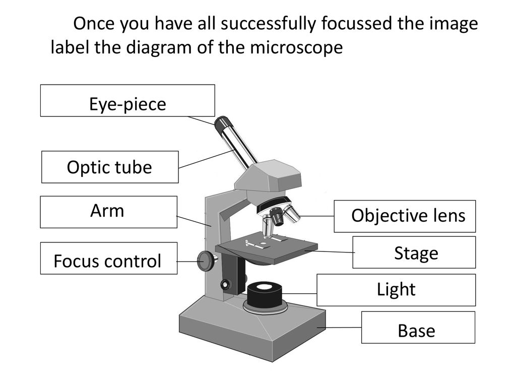
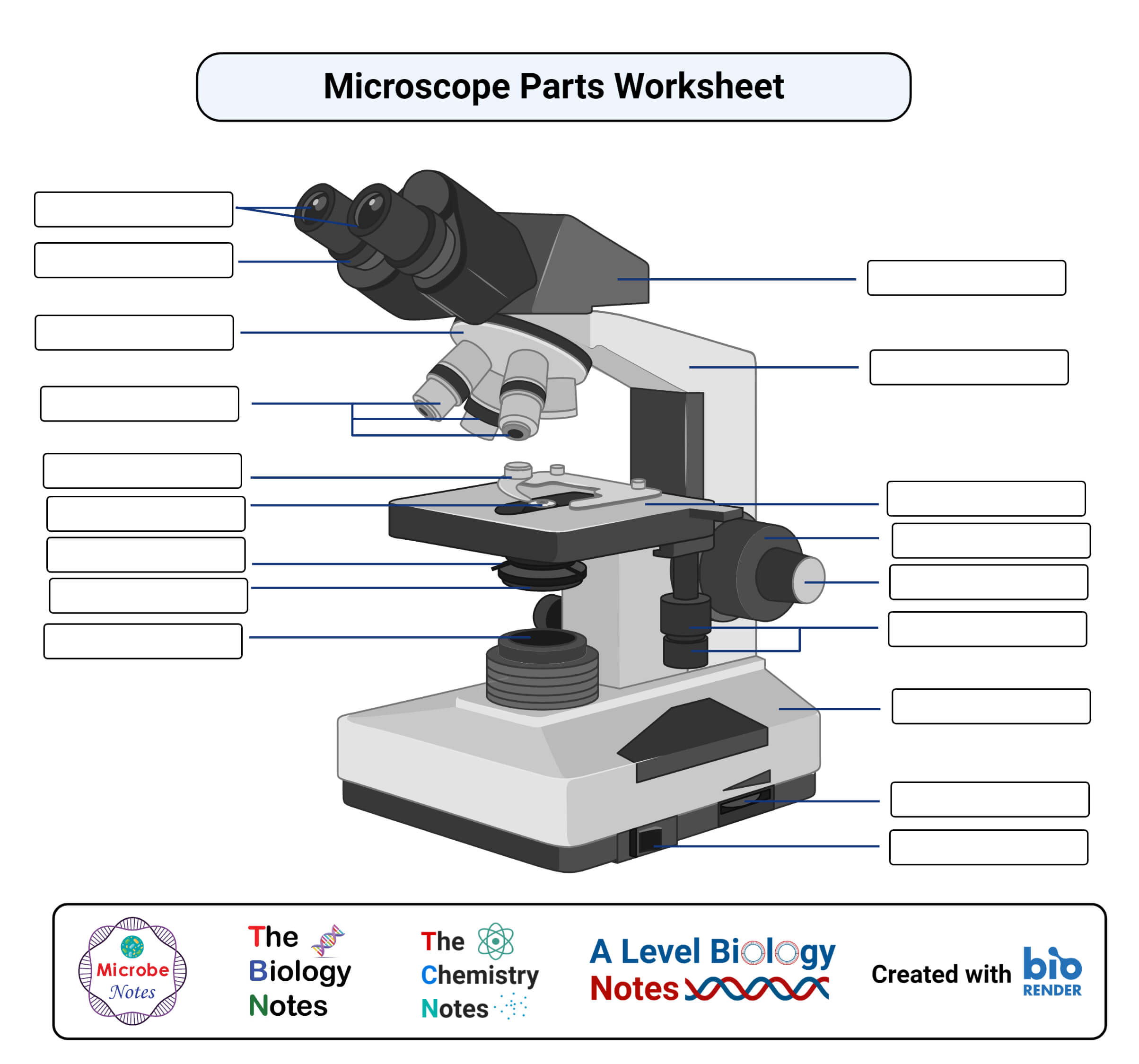
K To 12 Science Grade 7 Learners Material - Module ii. Science Grade 7 Learners Material First Edition, 2013 ISBN: _____ Republic Act 8293, section 176 states that: No copyright shall subsist in any work of the Government of the Philippines. However, prior approval of the government agency or office wherein the work is created shall be necessary for exploitation of such work for profit. ... Label A Microscope Teaching Resources | Teachers Pay Teachers The 13 parts of the microscope: microscope, base, arm, inclination joint, course adjustment, fine adjustment, body tube, ocular lens, revolving nose piece, objectives, stage, stage clips, and iris diaphragm.Includes:13 cards with labels13 cards without labels13 labels1 blackline masterCards with labels are approx. 3¾" x 4", cards without ... Label Microscope Diagram - EnchantedLearning.com Using the terms listed below, label the microscope diagram. arm - this attaches the eyepiece and body tube to the base. base - this supports the microscope. body tube - the tube that supports the eyepiece. coarse focus adjustment - a knob that makes large adjustments to the focus. diaphragm - an adjustable opening under the stage, allowing ... Microscope, Microscope Parts, Labeled Diagram, and Functions The description given below summarize the brief description of microscope parts used to visualize the microscopic specimens such as animal cells, plant cells, microbes, bacteria, viruses, microorganisms etc. The Microscopes parts divided into three different structural parts Head, Base, and Arms.
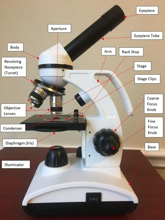
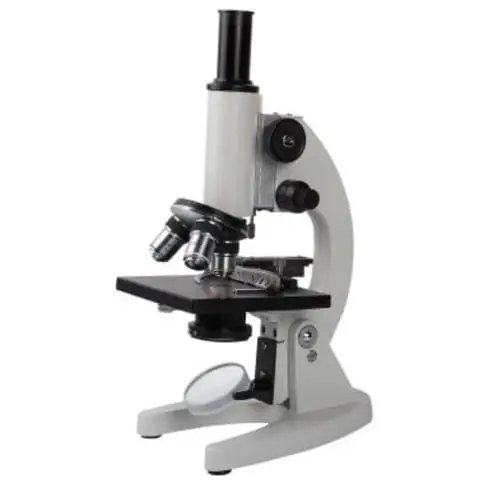
Microscope Parts and Functions A standard microscope has three, four, or five objective lenses that range in power from 4X to 100X. When focusing the microscope, be careful that the objective lens doesn't touch the slide, as it could break the slide and destroy the specimen. Specimen or slide: The specimen is the object being examined. Compound Microscope Parts - Labeled Diagram and their Functions The eyepiece (or ocular lens) is the lens part at the top of a microscope that the viewer looks through. The standard eyepiece has a magnification of 10x. You may exchange with an optional eyepiece ranging from 5x - 30x. [In this figure] The structure inside an eyepiece. The current design of the eyepiece is no longer a single convex lens. Parts of the Microscope with Labeling (also Free Printouts) A microscope is one of the invaluable tools in the laboratory setting. It is used to observe things that cannot be seen by the naked eye. Table of Contents, 1. Eyepiece, 2. Body tube/Head, 3. Turret/Nose piece, 4. Objective lenses, 5. Knobs (fine and coarse) 6. Stage and stage clips, 7. Aperture, 9. Condenser, 10. Condenser focus knob, 11. en.wikipedia.org › wiki › Electron_microscopeElectron microscope - Wikipedia An electron microscope is a microscope that uses a beam of accelerated electrons as a source of illumination. As the wavelength of an electron can be up to 100,000 times shorter than that of visible light photons , electron microscopes have a higher resolving power than light microscopes and can reveal the structure of smaller objects.
Compound Light Microscope Labeling Diagram | Quizlet
Labelled Diagram of Compound Microscope The below mentioned article provides a labelled diagram of compound microscope. Part # 1. The Stand: The stand is made up of a heavy foot which carries a curved inclinable limb or arm bearing the body tube. The foot is generally horse shoe-shaped structure (Fig. 2) which rests on table top or any other surface on which the microscope in kept.

Labeled Parts Of A Microscope - ClipArt Best
Parts of a microscope with functions and labeled diagram - Microbe Notes Figure: Diagram of parts of a microscope, There are three structural parts of the microscope i.e. head, base, and arm. Head - This is also known as the body. It carries the optical parts in the upper part of the microscope. Base - It acts as microscopes support. It also carries microscopic illuminators.
16 Parts of a Compound Microscope: Diagrams and Video ...
brain diagram without labels Download Female Shadow Anatomy Without Labels - Human Body Without. 11 Images about Download Female Shadow Anatomy Without Labels - Human Body Without : Fill In The Blank Brain Diagram Blank Brain Diagram Coloring Pages, Бульбарный паралич: диагностика, симптомы и лечение and also Human Skull No Text No Color Clip Art at Clker.com - vector clip art.
Microscope labelling 11 - Teaching resources
Microscope Labeling Game - PurposeGames.com About this Quiz. This is an online quiz called Microscope Labeling Game. There is a printable worksheet available for download here so you can take the quiz with pen and paper. This quiz has tags. Click on the tags below to find other quizzes on the same subject. Science.

Compound Microscope Parts – Labeled Diagram and their ...
Nuclear envelope - Wikipedia The nuclear envelope is punctured by around a thousand nuclear pore complexes, about 100 nm across, with an inner channel about 40 nm wide. The complexes contain a number of nucleoporins, proteins that link the inner and outer nuclear membranes.. Cell division. During the G2 phase of interphase, the nuclear membrane increases its surface area and doubles its …

Parts of the Microscope with Labeling (also Free Printouts ...
Compound Microscope Parts, Functions, and Labeled Diagram Compound Microscope Definitions for Labels. Eyepiece (ocular lens) with or without Pointer: The part that is looked through at the top of the compound microscope. Eyepieces typically have a magnification between 5x & 30x. Monocular or Binocular Head: Structural support that holds & connects the eyepieces to the objective lenses.

How to use a Microscope - Microscopes 4 Schools
Microscope Label Interactive Worksheets & Teaching Resources | TpT Microscope Interactive Notebook Activity, by, Jodi's Jewels, 12, $1.89, PDF, Students will complete a timeline of the history of the microscope, label a diagram, and create a pocket foldable with terms and definition cards. The timeline can be completed according to the teacher's directions or like the answer key example.
Parts of a Microscope Quiz
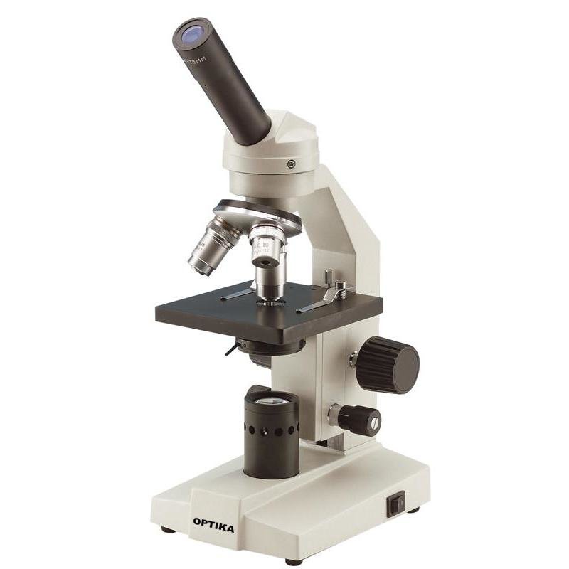
Simple Microscope - Parts, Functions, Diagram and Labelling A simple microscope is a device that only has one lens for magnification. It functions the same way as the magnifying glass. Although it is simple in terms of design and function, it is useful I various fields including medicine, jewelry and watchmaking, and agriculture, to name a few. References, ,
Parts of a Light Microscope Activity | Labeling Task
› searchImages, Stock Photos & Vectors | Shutterstock Jun 30, 2022 · Find stock images in HD and millions of other royalty-free stock photos, illustrations and vectors in the Shutterstock collection. Thousands of new, high-quality pictures added every day.
Compound Microscope Parts, Functions, and Labeled Diagram ...
rockyourhomeschool.net › microscope-worksheetsFree Microscope Worksheets for Simple Science Fun for Your ... Parts of a Microscope . The first worksheet labels the different parts of a microscope, including the base, slide holder, and condenser. If you have a microscope, compare and contrast this worksheet to it. Also, your kids can color this microscope diagram in and read the words to each part of the microscope.
Free Microscope Drawing, Download Free Microscope Drawing png ...
The Contrast Transfer Function - Image Formation | Coursera 14.12.2019 · The violins, without a microphone, would be like a position here in spatial frequency where you don't hear them at all. So contrast transfer function tells us how much of each of these components are going to arrive at the image. Just like placing the microphones in different positions in an orchestra tells you which of the instruments we hear strongly and which will be …
Compound Microscope - Types, Parts, Diagram, Functions and ...
Compound Microscope: Definition, Diagram, Parts, Uses, Working ... - BYJUS The compound microscope is mainly used for studying the structural details of cell, tissue, or sections of organs. The parts of a compound microscope can be classified into two: Non-optical parts, Optical parts, Non-optical parts, Base, The base is also known as the foot which is either U or horseshoe-shaped.

Microscope Labeling Diagram | Quizlet
Fluorescence Resonance Energy Transfer (FRET) Microscopy Microscope configurational parameters for fluorescence resonance energy transfer investigations vary with the requirements of the fluorophores, specimen, and imaging mode(s), but virtually any upright or inverted microscope can be retrofitted for FRET microscopy (see Figure 7). In general, the microscope should be equipped with a high-resolution (12-bit) cooled and …

Parts of a microscope with functions and labeled diagram
Electron microscope - Wikipedia An electron microscope is a microscope that uses a beam of accelerated electrons as a source of illumination. As the wavelength of an electron can be up to 100,000 times shorter than that of visible light photons, electron microscopes have a higher resolving power than light microscopes and can reveal the structure of smaller objects.. Electron microscopes use shaped magnetic …

Labeling a Microscope Free Worksheet Pack
19+ Animal Cell Diagram Without Labels Best A simple diagram of an unspecialised animal cell without labels. The most important structures of plant and animal cells are shown in the diagrams below, which provide a clear illustration of how much these cells have in common. The first is a colored and labeled cell diagram. Cells are covered by a cell membrane and come in many different shapes.
Microscope Using a microscope, I have developed my ...
Label the microscope — Science Learning Hub In this interactive, you can label the different parts of a microscope. Use this with the Microscope parts activity to help students identify and label the main parts of a microscope and then describe their functions. Drag and drop the text labels onto the microscope diagram.
How to Use the Microscope
PDF Parts of a Microscope Printables - Homeschool Creations What are the three magnification levels on a microscope's objective lenses? 4x, 10x, and 40x. The eyepiece on a microscope magnifies at 10x, so when used together, the 4x lens magnifies an item 40x, the 10x magnifies 100x, and the 40x magnifies 400x. (note: for typical student microscope -other microscopes will vary)

Unlabeled Microscope Diagram posted by Ryan Mercado
Diagram of a Compound Microscope - Biology Discussion 1. It is noted first that which objective lens is in use on the microscope. 2. Stage micrometer is positioned in such a way that it is in the field of view. 3. The eyepiece is rotated so that the two scales, the eyepiece or ocular scale and the stage micrometer scale, are parallel. 4.
Parts of a microscope with functions and labeled diagram
Images, Stock Photos & Vectors | Shutterstock 30.06.2022 · Find stock images in HD and millions of other royalty-free stock photos, illustrations and vectors in the Shutterstock collection. Thousands of new, high-quality pictures added every day.
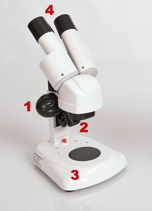
Compound and Stereo- microscopes - Microscopes 4 Schools
Free Microscope Worksheets for Simple Science Fun for Your Students 1. Parts of a Microscope . The first worksheet labels the different parts of a microscope, including the base, slide holder, and condenser. If you have a microscope, compare and contrast this worksheet to it.Also, your kids can color this microscope diagram in and read the words to each part of the microscope.

Biology Microscope Labeling and Definitions (Light/Compound ...
rsscience.com › stereo-microscopeParts of Stereo Microscope (Dissecting microscope) – labeled ... Labeled part diagram of a stereo microscope Major structural parts of a stereo microscope. There are three major structural parts of a stereo microscope. The viewing Head includes the upper part of the microscope, which houses the most critical optical components, including the eyepiece, objective lens, and light source of the microscope.
How to see a plant cell under a compound microscope - Quora
ALEX | Alabama Learning Exchange Subject: Digital Literacy and Computer Science (4), Science (4) Title: Using Code to Create an Animated Animal Description: Students will use the free online coding program, Scratch, to learn the basics of coding and how to use blocks and animations to create an animated animal. Students will show how an animated animal will receive, process, and respond to information …

Parts of a Compound Microscope and Their Functions
Label the microscope Diagram | Quizlet Label the microscope Diagram | Quizlet, Label the microscope, 3.8 (12 reviews) + −, Flashcards, Learn, Test, Match, Created by, Rdavis952 TEACHER, Terms in this set (14) Ocular, eye piece, Nosepiece, Holds the objectives and can be rotated to change the magnification, 10x Objective, Medium power, 4x objective, Low Power, 40x objective, high power,

How to draw compound of Microscope easily - step by step
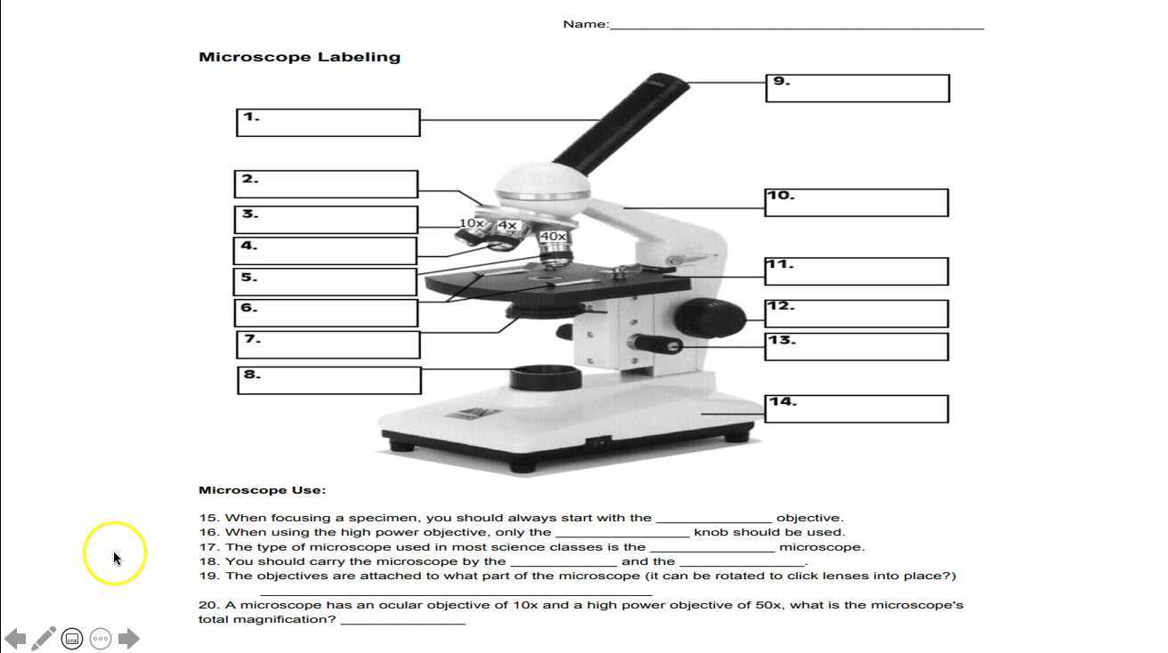
Microscope Labeling - The Biology Corner Students label the parts of the microscope in this photo of a basic laboratory light microscope. Can be used for practice or as a quiz. ... The type of microscope used in most science classes is the _____ microscope. 18. You should carry the microscope by the _____ and the _____. 19. The objectives are attached to what part of the microscope ...
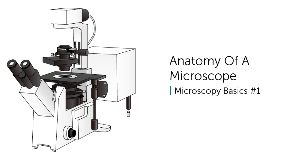
Anatomy Of A Microscope
16 Parts of a Compound Microscope: Diagrams and Video Once you have an understanding of the parts of the microscope it will be much easier to navigate around and begin observing your specimen, which is the fun part! The 16 core parts of a compound microscope are: Head (Body) Arm, Base, Eyepiece, Eyepiece tube, Objective lenses, Revolving Nosepiece (Turret) Rack stop, Coarse adjustment knobs,

Compound Microscope Parts, Functions, and Labeled Diagram ...
Microscope Labeling Activity | Sticky Note Labels - Classful This clear and simple microscope labeling activity is exactly what your students need to learn the parts of a compound microscope! There are five versions of microscope labeling diagrams included: ⭐Sticky Note microscope to label ⭐Microscope to label with a word bank ⭐Microscope to label without a word bank ⭐Plain microscope to label without lines ⭐Version with and without name/date ...

Microscope Diagram - Label Diagram | Quizlet
7th grade Science - Microscope Diagram | Quizlet The Parts of a Microscope. 12 terms. totobear PLUS. Sets found in the same folder. Science Key terms 7th grade. 13 terms. palocastillo. 7th Grade Earth Science. 9 terms. EliseC17. 7thGrade Review - Cells/Biology. 26 terms. SolizScience TEACHER. 7th grade Science, Cell theory. 8 terms. Super1412. Other sets by this creator.
The Compound Light Microscope Label the following parts on ...
Label the Microscope Diagram | Download Scientific Diagram - ResearchGate Download scientific diagram | Label the Microscope Diagram from publication: Laboratory Exercises in Microbiology: Discovering the Unseen World through Hands-on Investigation | Microbiology ...

Microscope labeled diagram
Microscope Objective Lens | Products | Leica Microsystems Microscope Objectives. Leica Microsystems – The Ultimate in Optical Competence. For more than 170 years Leica Microsystems has designed and produced top-class objectives for a wide variety of applications in research, industry and medicine. The optics specialists at Leica Microsystems bring the highest level of experience and expertise to bear in reducing …
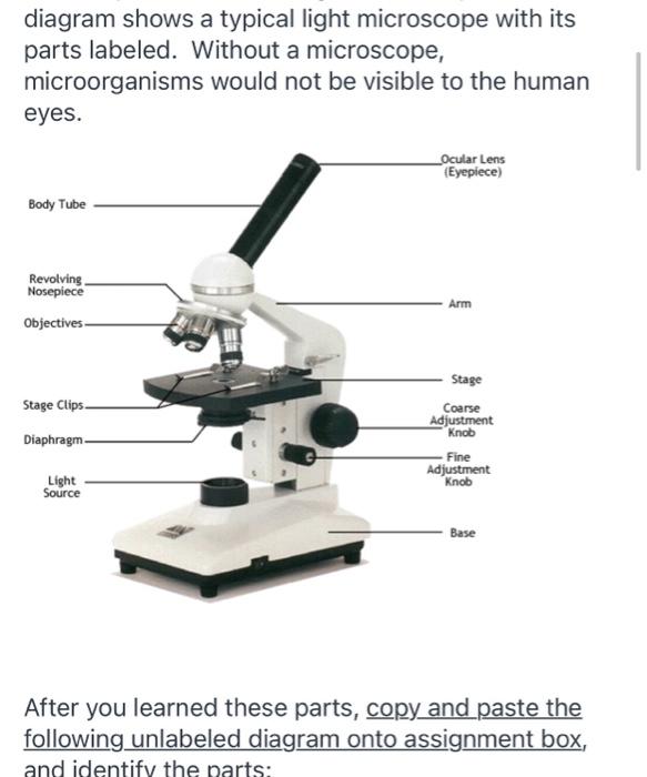
Solved diagram shows a typical light microscope with its ...

Compound Microscope- Definition, Labeled Diagram, Principle ...

Light Microscope- Definition, Principle, Types, Parts ...

Label the Microscope Diagram | Quizlet

Introduction to Microscopes - ppt download

Label the Microscope Diagram | Download Scientific Diagram

Microscope Diagram Labeled, Unlabeled and Blank | Parts of a ...

Label a Microscope Worksheet
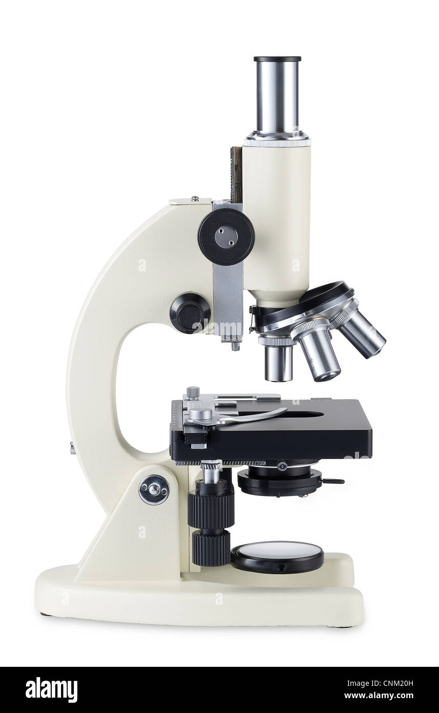
Microscope hi-res stock photography and images - Alamy

Label the microscope — Science Learning Hub
Microscope Labeling

Compound Microscope: Parts of Compound Microscope
Post a Comment for "41 microscope diagram without labels"